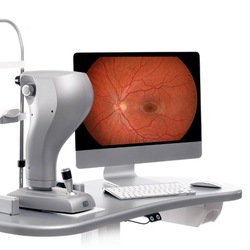
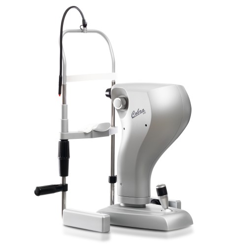
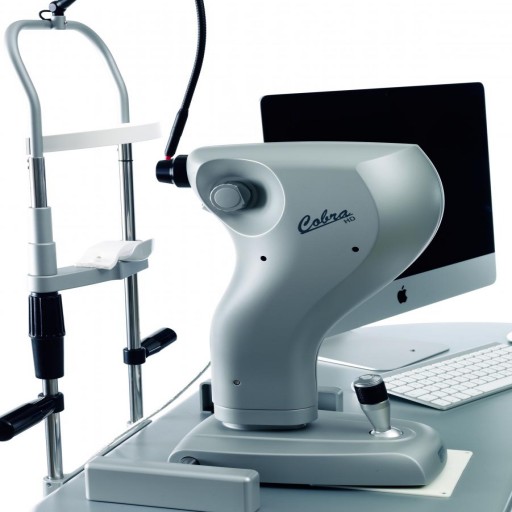


Cobra HD is a non-mydriatic digital fundus camera that comprises all the functions required for a rapid screening of the status of the retina. Cobra uses an innovative optical system that can provide high quality images of the ocular fundus. With its ergonomic design Cobra provides a clear and detailed image of the ocular fundus with a field of vision of up to 50 degrees. Cobra uses a minimum flash exposure, allowing a fast and detailed acquisition of the fundus and minimizing the discomfort for the patient. Cobra HD shares the use of the CCD high-resolution sensor (5 megapixel) for the alignment of the pa- tient (with IR illumination) and the capture of retinal images (with a white light flash and IR LEDs).
- 50 Degree field of view.
- Offers retinal photography with minimum flash exposure allowing quick and efficient fundus photography, thereby minimizing patient discomfort.
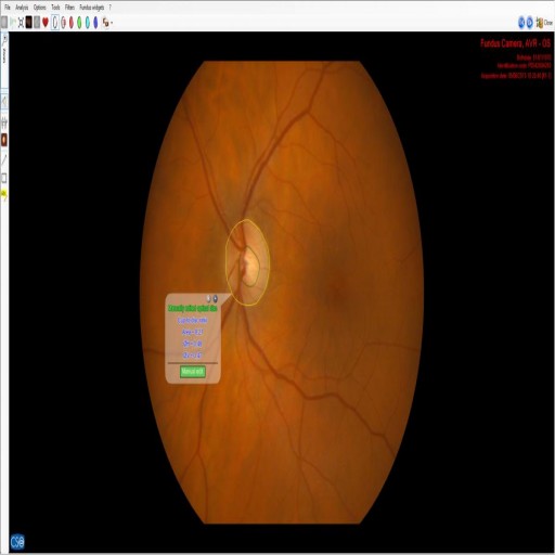
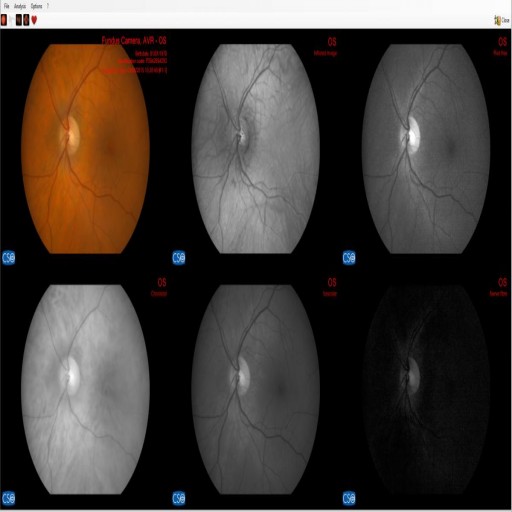
- Multiple wave-length images can be displayed on one screen: the original image, infrared image, red-free image, as well the choroidal, vascular and nerve fiber image.
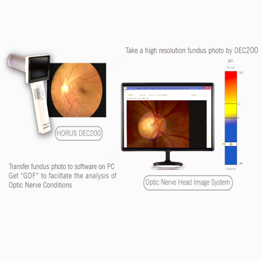
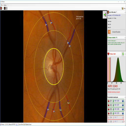
- The measurement of the Cup to Disk ratio is easily acheived using the built in measurement tools that are available in the Phoenix software platform for the detection of glaucomatous disease.
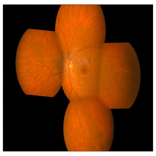
- Cobra HD allows the acquisition of multiple images, to create a panoramic image of the peripheral retinal areas.
- Cobra HD includes a module for the analysis of the Meibomian Glands (MGD). Using Pheonix software, the glands structure and health can be analysed.
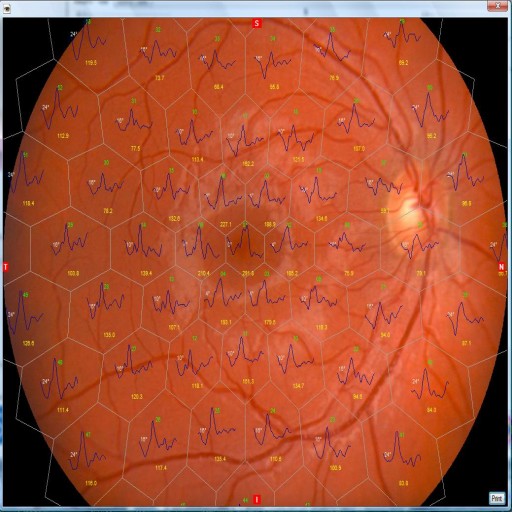
- The image of the retinal fundus provided by COBRA can be combined with the multifocal ERG test, performed with the RETIMAX device. This new module provides a precise indication of the functionality of every analyzed retinal area; it is very useful for the diagnosis and the follow-up of Macular Degeneration as well as degenerative hereditary retinal diseases. *optional module
- The AVR tool(Optional) measures the relationship between the branch arteriolar-venous diameter. A low relationship between the dimension of the vessels, may be predictive of cardiovascular problems in adult patients.
- USB connection enables a fast, easy transfer, and saving images
- Compatible with DICOM.
- Image processing, drawing and measuring.
- C/D ratio, Disc HV, Cup HV
- Drawing (text/objects can be inserted)
- Very light weight of 6 KG.
- 2 years Exclusive warranty from manufacturer.
Brochures
hello.pdfMultimedia
Request a Quote
Please fill out the form below to request a quote. We will be in contact with you shortly.