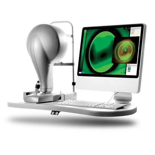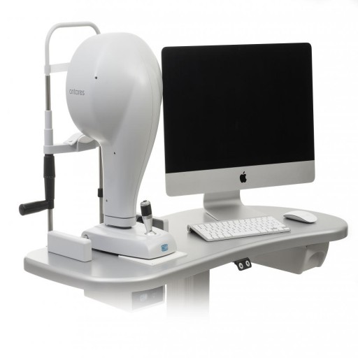
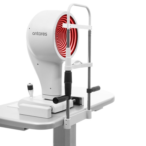
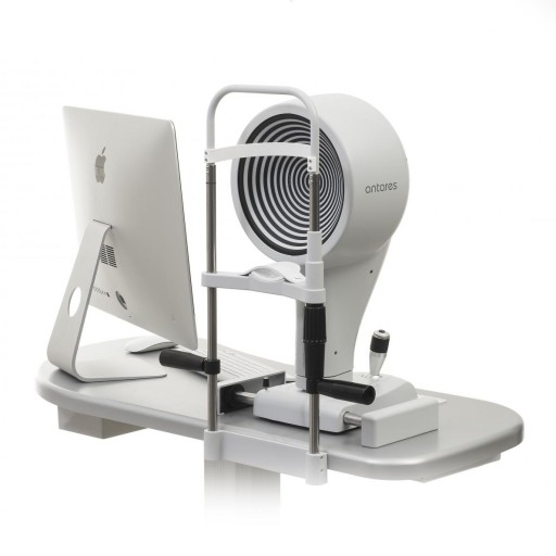
ANTARES is a medical device used to perform the corneal topography and for the diagnostic of the lacrimal dysfunction.The device has been designed for the capture and the elaboration of an image of the cornea in the ophthalmic procedure.
Thanks to the high-resolution colour video camera, you can film "live" the corneal surface and visualize it on the computer screen.
The device provides information on the curvature, the elevation and the refractive power, and a great number of concise parameters for the diagnostic and the follow-up of the corneal surface.
Extensive functions such as Corneal topography,Pupillography,Meibography,Analysis of the tear film,Videokeratoscopy,Keratoconus screening,Module for contact lenses application and comprehensive Dry Eye Report are available.
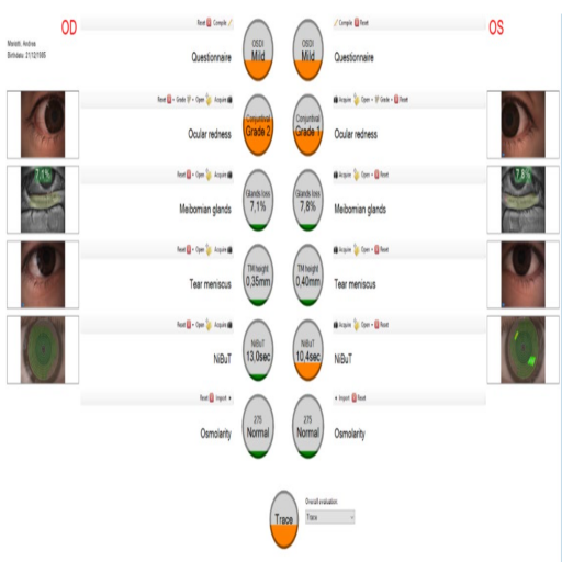
Based on the Ocular Surface Disease Index questionnaire (OSDI), limbal and conjunctival hyperaemia, Meibomian glands analysis, tear meniscus analysis, NIBUT, and tear osmolarity, calculated merging together all partial scores, provides an overall evaluation of the clinical condition of the patient for a comprehesive diagnosis of the dry eye disease.
DRY EYE REPORT
The Dry Eye Report (DER) is a collection of 5 examinations and a questionnaire (OSDI) assigned to a single patient.The main purpose of the report is to summarize all available examinations concerning the dry eye condition and to ease its diagnosis.With all examination previews and gradings collected together in a single page, it is much easier to get an overview of the patient's condition.Eventually a single DER can be graded by the clinic with an overall score ranging from Normal to Severe.
FEATURES DRY EYE REPORT:
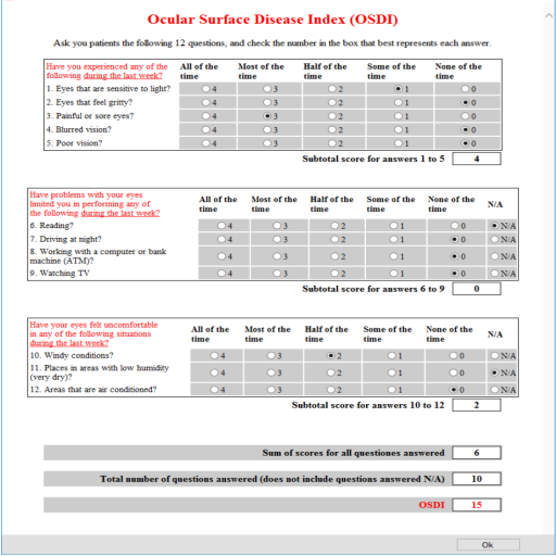
- The Ocular Surface Disease Index (OSDI) that is a standardized questionnaire developed for measuring the severity of DED. It is answered by the patient before any other examination is performed.
- Ocular redness (or conjunctival Hyperaemia according to original Efron definition) analysis: a limbal and/or conjuntival picture of the eye is acquired and manually graded with the help of a reference grading scale.
- Meibomian glands analysis: a picture of Meibomian glands is acquired through infrared illumination and is further classified with the assistance of a specific software tool.
- Tear meniscus analysis: it is widely used in clinical practice and shows good correlation with other DED tests. A calibrated picture of the tear meniscus is acquired and then is manually marked to measure the mean tear meniscus height (TMH) and its standard deviation using a specific software tool.
- NIBUT: requires the keratoscope, scheimpflug camera or AS-OCT NiBut examination. The projected Placido disc ring pattern is recorded as a movie and analysed automatically by the system. Then the score is computed by evaluating the First Break-Up time in the rings structure.
- Osmolarity - Input from external device can be used for final report.
Brochures
hello.pdfMultimedia
Request a Quote
Please fill out the form below to request a quote. We will be in contact with you shortly.